
Endodontics & Tooth Pain Guide
About Your Tooth
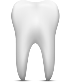 Your tooth consists of two main parts: the crown, which is that part of the tooth above the gum and visible in your mouth; and the root or roots, which is that part of the tooth that lies beneath the gum and is surrounded by bone. Inside each root is a channel that runs the length of the tooth. This channel is the root canal and contains the pulp (nerves, blood vessels, and soft tissue), which is often referred to as the “nerve” of the tooth. The pulp may be irreversibly damaged by bacteria associated with decay, very deep restorations, fractures, trauma, or periodontal disease.
In order to preserve a tooth in which this has occurred, it is necessary to remove the diseased pulp tissue. This procedure is known as endodontic therapy. Since endodontic therapy is concerned with removing only the pulp from the root canal, the root will continue to function normally because the supporting tissues remain intact. It is advisable to remove the injured pulp because it may become infected or act as an irritant to the tissues surrounding the tooth.
Want To Learn More?
Our caring staff is here to help you if you have any questions!
Tooth Pain Guide
Please select from the list below the title that best reflects your pain.
Momentary sensitivity to hot or cold foods
Sensitivity to hot or cold foods after dental treatment
Sharp pain when biting down on food
Lingering pain after eating hot or cold foods
Constant and severe pain and pressure, swelling of gum, and sensitivity to touch
Dull ache and pressure in upper teeth and jaw
Chronic pain in head, neck, or ear
Want To Learn More?
Our caring staff is here to help you if you have any questions!
>
Endodontic FAQ
What is endodontics?
Endodontics is a branch of dentistry recognized by the American Dental Association involving treatment of the pulp (root canal) and surrounding tissues of the tooth. When you look at your tooth in the mirror, what you see is the crown. The rest of the tooth, the portion hidden beneath the gum line, is called the root. Though the outer portion of the root is a hard tissue called dentin, the inside channel or “root canal” contains a pulp of soft tissue, blood vessels and nerves. Bacteria that are introduced into the pulp as a result of tooth decay, periodontal disease, tooth fracture or other problems, can severely damage the pulp. When that happens, an endodontic specialist removes the diseased pulp to save the tooth and prevent further infection and inflammation. After successful endodontic treatment, the tooth continues to perform normally.
<
I’m worried about x-rays. Should I be?
No. While x-rays will be necessary during your endodontics treatment, we use an advanced non-film computerized system, called digital radiography, that produces radiation levels up to 90 percent lower than those of already low dose conventional dental x-ray machinery. These digital images can be optimized, archived, printed and sent to cotherapists via e-mail or CD-ROM. For more information contact Sirona Dental Systems, Inc.
What about infection?
Again, there’s no need for concern. We adhere to the most rigorous standards of infection control advocated by OSHA, the Centers for Disease Control and the American Dental Association. We utilize autoclave sterilization and barrier techniques to eliminate any risk of infection.
What happens after treatment?
When your root canal therapy has been completed, a record of your treatment will be sent to your restorative dentist. You should contact his office for a follow-up restoration within a few weeks of completion at our office. Your restorative dentist will decide on what type of restoration is necessary to protect your tooth. It is rare for endodontic patients to experience complications after routine endodontic treatment or microsurgery. If a problem does occur, however, we are available at all times to respond.
What new technologies are being used?
Operating Microscopes:
In addition to digital radiography, we utilize special Carl Zeiss operating microscopes. Magnification and fiber optic illumination are helpful in aiding the doctor to see tiny details inside your tooth. Also, a tiny video camera on the operating microscope can record images of your tooth to further document the doctor’s findings.
Cone Beam Computed Tomography (CBCT) 3D X-ray machine:
See the Advanced Technology page.
Want To Learn More?
Our caring staff is here to help you if you have any questions!